EFFECT OF MECHANICAL MINCING ON MINIMALLY MANIPULATED ARTICULAR CARTILAGE FOR SURGICAL TRANSPLANTATION
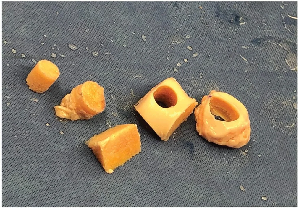
Figure 1 - Femoral condylar allograft after osteochondral allograft removal.
Background:
Point-of-care treatment options for medium to large symptomatic articular cartilage defects are limited. Minced cartilage implantation is an encouraging single-stage option, providing fresh viable autologous tissue with minimal morbidity and cost.
Purpose:
To determine the histological properties of mechanically minced versus minimally manipulated articular cartilage.
Study Design:
Controlled laboratory study.
Methods:
Remnant articular cartilage was collected from fresh femoral condylar allografts. Cartilage samples were divided into 4 groups: cartilage explants with or without fibrin glue and mechanically minced cartilage with or without fibrin glue. Samples were cultured for 42 days. Chondrocyte viability was assessed using live/dead assay. Cellular migration and outgrowth were monitored using bright-field microscopy. Extracellular matrix deposition was assessed via histological staining. Proteoglycan content and synthesis were assessed using dimethylmethylene blue assay and radiolabeled 35S-sulfate, respectively. Type II collagen (COL2A1) gene expression was analyzed via polymerase chain reaction.
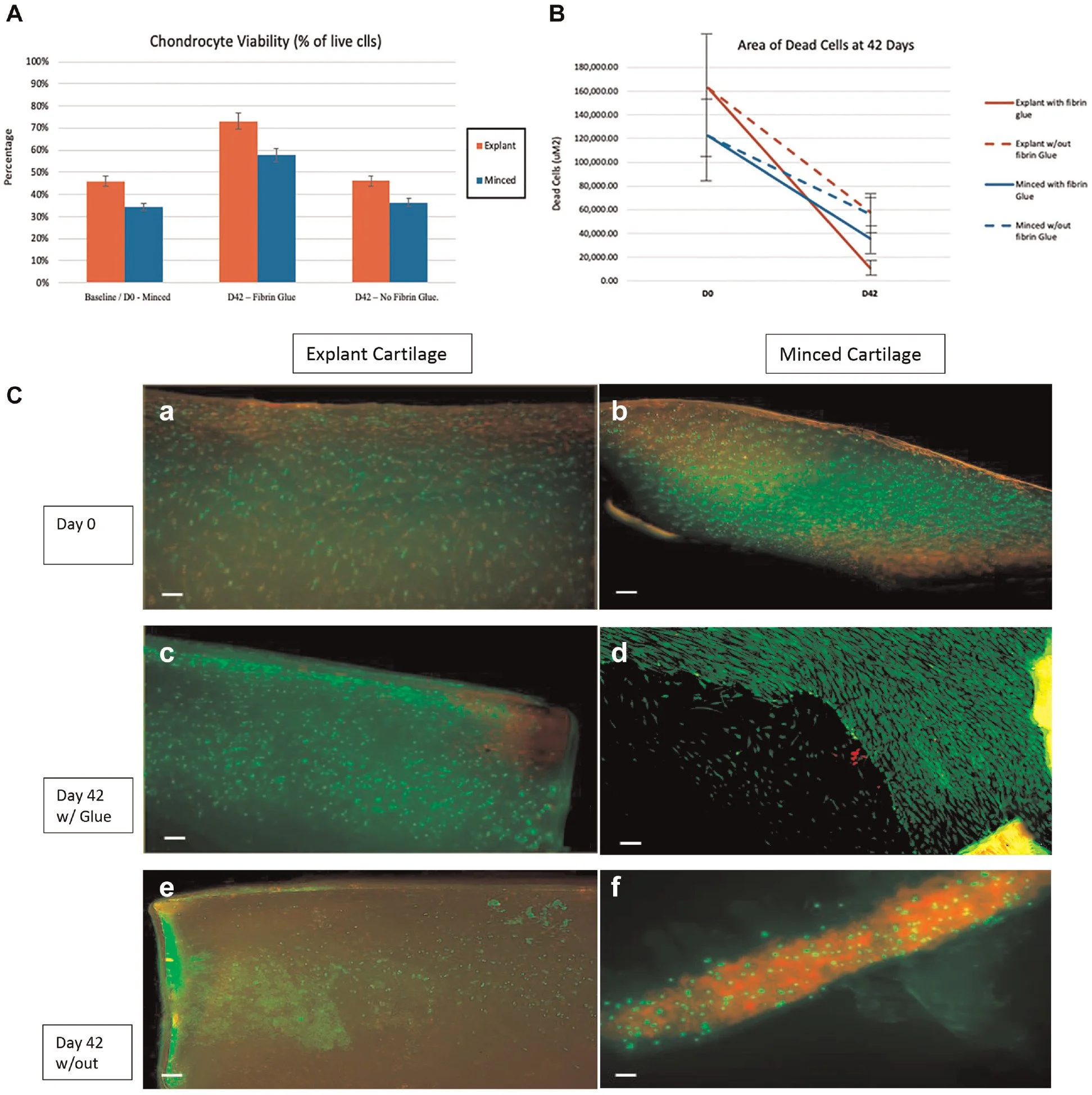
Figure 2 - Effect of mincing on chondrocyte viability. (A) Comparison with baseline of chondrocyte viability for explant and minced cartilage on day 0 (D0) and day 42 (D42). (B) Area of cell death on day 0 and day 42. (C) Live/dead staining of minced and explant cartilage with and without fibrin glue. Representative images: explant cartilage on day 0 (a), minced cartilage on day 0 (b), explant cartilage with glue on day 42 (c), minced cartilage with glue on day 42 (d), explant cartilage without glue on day 42 (e), and minced cartilage without glue on day 42 (f). Scale bars: 100 μm.
Results:
The mean viability of minced cartilage particles (34% ± 14%) was not significantly reduced compared with baseline (46% ± 13%) on day 0 (P = .90). After culture, no significant difference in the percentage of live cells was appreciated between mechanically minced (58% ± 23%) and explant (73% ± 14%) cartilage in the presence of fibrin glue (P = .52). The addition of fibrin glue did not significantly affect the viability of cartilage samples. The qualitative assessment revealed comparable cellular migration and outgrowth between groups. Proteoglycan synthesis was not significantly different between groups. Histological analysis findings were positive for COL2A1 in all groups, and matrix formation was appreciated in all groups. COL2A1 expression in minced cartilage (1.72 ± 1.88) was significantly higher than in explant cartilage (0.15 ± 0.07) in the presence of fibrin glue (P = .01).
Conclusion:
Mechanically minced articular cartilage remained viable after 42 days of culture in vitro and was comparable with cartilage explants with regard to cellular migration, outgrowth, and extracellular matrix synthesis.
Click on the link for the full print article:
Published June 23, 2022 in the American Journal of Sports Medicine (Volume 50 - Issue 9).


